Diagram of a Horse Hoof Detailed Cross Section
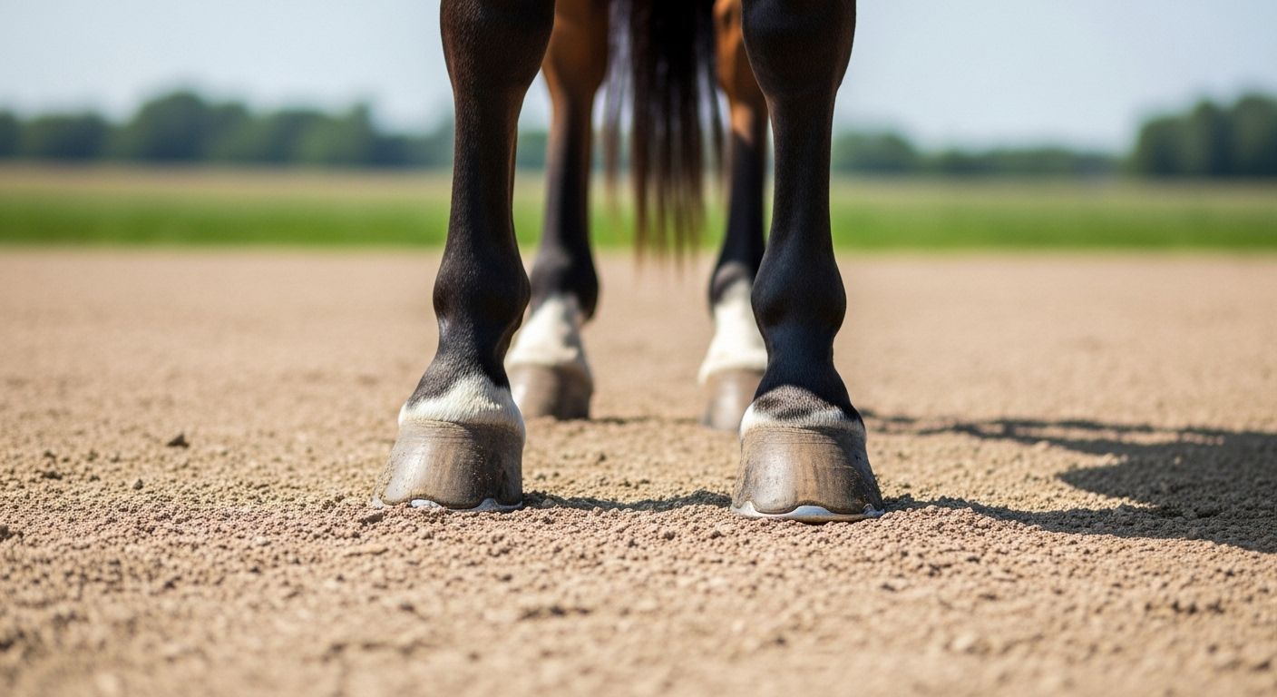
Table of Contents
diagram of a horse hoof
What’s in a Hoof? More Than Just a Fancy Shoe Holder
Ever stared at a horse’s hoof and thought, *“Blimey, that’s just a big toenail, innit?”*—mate, that’s like callin’ Big Ben “a posh clock tower”. The diagram of a horse hoof is less *manicure* and more *mechanical marvel*: shock absorber, blood pump, sensory hub, and load distributor—all wrapped in keratin tougher than yer nan’s Christmas pudding. One hoof bears ~250 kg per stride at trot—equivalent to jumpin’ off a garden wall *every other second*. And yet, it *expands*, *contracts*, *breathes*. Get the diagram of a horse hoof right in yer head, and you’ll never look at a farrier’s rasp the same way again.
What Is the Anatomy of a Horse’s Hoof? Let’s Crack It Open Like a Quality Scotch Egg
Right—peel back the shell, and here’s what the diagram of a horse hoof really holds. Externally: *wall*, *sole*, *frog*, *heel bulbs*, *white line*. Internally? Where the magic (and misery) lives:
- Distal phalanx (P3 / coffin bone) – the keystone. Suspended by 600+ laminae. Tip it 5°, and the whole house groans.
- Navicular bone – tiny sesamoid, massive influence. Acts as a pulley for the deep digital flexor tendon (DDFT). Wear it down, and every step’s agony.
- Digital cushion – that fibro-fatty pad behind the frog. Not bone, but *critical*. Compresses on impact, pumps blood back up the leg like a heart in trainers.
- Laminae – the Velcro between bone and hoof wall. Fail here? Hello, laminitis.
A healthy hoof’s not just *hard*—it’s *dynamic*. The diagram of a horse hoof isn’t a static sketch; it’s a live blueprint of biomechanics in motion. Miss one piece, and the whole symphony goes flat.
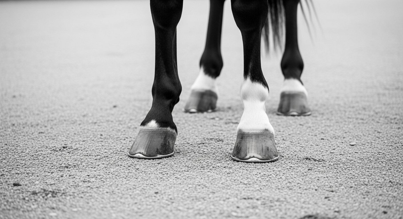
How Should a Horse’s Hoof Look? Not “Pretty”—*Functional*.
Forget Instagram symmetry—what matters is *physiology*, not aesthetics. In a proper diagram of a horse hoof, balance isn’t about matching angles; it’s about *load distribution*:
- Wall thickness: 6–12 mm at toe, tapering slightly—thinner than a crisp packet, stronger than steel (per gram).
- Frog: broad, elastic, ground-contacting—*not* recessed like a shy hedgehog. Should leave a print in soft ground, every time.
- Heel height: level with the frog’s base—no “underrun” heels (where the heel curls forward like a bad perm).
- White line: tight, narrow (<2 mm), *not* crumbly or stretched.
Fun stat? A 500 kg horse’s hoof has ~60 cm² of ground contact—less than *two A4 sheets*. Yet it handles ~1,000 kg of peak force at gallop. That’s why the diagram of a horse hoof must reflect *function first, fashion never*.
The Middle Part of a Horse’s Hoof? It’s Not the Frog—It’s the *Solar Surface*
“Middle part?”—ah, the classic pub-quiz trap. Most’ll shout *frog*, but the *true* central zone (in ground-contact terms) is the solar surface—the *sole*, *frog*, and *bars* working as one unit. In the diagram of a horse hoof, this trio forms the “tripod” of support:
- Sole: concave, protective—*not* weight-bearing unless pathological.
- Frog: the shock absorber & circulator—*must* touch ground for digital cushion activation.
- Bars: inward folds of the wall—stabilise the heel, prevent over-expansion.
When the farrier says, *“He’s not loading the back of the foot,”* they mean the frog’s *floating*—a silent red flag. Because in the diagram of a horse hoof, the middle isn’t a *point*—it’s a *system*.
Foot or Hoof? Semantics, Love—But Biomechanically, It’s a *Single Toe*
“Does a horse have a foot or a hoof?”—technically? It’s *both*, but let’s split hairs like a pedantic don. “Foot” = the *entire distal limb* (pastern down). “Hoof” = the *keratinised capsule*—the bit that *click-clacks* on tarmac. But evolutionarily? It’s one *hyper-specialised digit*—digit III, to be precise. The other toes? Gone. Vanished. *Poof*. Left behind two tiny splint bones (MCII/MTII & MCIV/MTIV)—vestigial ghosts in the diagram of a horse hoof. So yes—he’s got a *foot*, but he walks on *one toe*, encased in the toughest biological composite known to man. Call it a hoof. Call it a foot. Just don’t call it *simple*.
The Hoof as a Hydraulic Pump: How Blood Gets Back Uphill (Without a Lift)
Digital Cushion + Frog = The Heart’s Helper
Here’s the kicker most riders miss: the hoof *pumps blood*. Every time it loads, the diagram of a horse hoof springs into action—frog compresses, digital cushion squishes, venous plexuses empty toward the heart. Unload? It rebounds, sucking fresh blood in. This “hoof mechanism” contributes up to *30%* of venous return from the limb. No movement = poor circulation = colder feet = higher laminitis risk. That’s why box rest’s risky, and why turnout’s non-negotiable. The diagram of a horse hoof isn’t just *structure*—it’s *physiology in motion*.
Common Hoof Pathologies—And What the Diagram Shows First
Before the limp, before the heat—*the diagram whispers*. In the diagram of a horse hoof, early signs hide in plain sight:
| Visual Clue | Potential Issue | Action Window |
|---|---|---|
| Stretched white line, divergent growth rings | Laminae stress / subclinical laminitis🟢 2–4 weeks | |
| Contracted heels, recessed frog | Underrun heels, poor digital cushion development | 🟡 4–8 weeks |
| Asymmetrical wear, medial-lateral imbalance | Conformation strain or old injury compensation | 🟢 Ongoing |
| Deep central sulcus crack, foul odour | Thrush + possible frog prolapse | 🔴 <7 days |
Spot it in the diagram of a horse hoof? You’ve bought time. Ignore it? You’re ringing the vet *and* the remedial farrier—£180/hour, love.
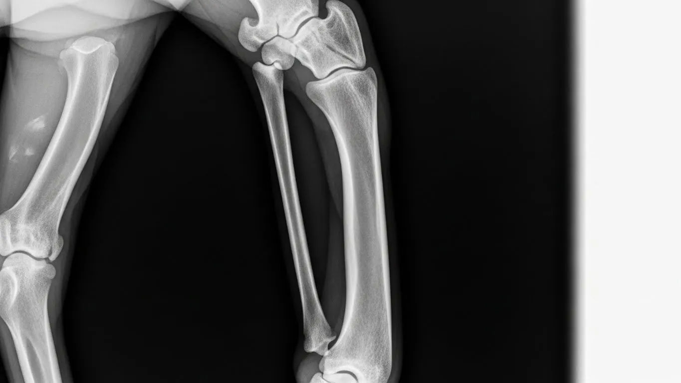
Trimming & Shoeing: Reading the Diagram Like a GP Reads an X-Ray
Trimming isn’t *sculpting*—it’s *rebalancing physics*. A good farrier uses the diagram of a horse hoof as a guide:
- Breakover: where the toe *leaves* the ground. Too long? Strain on DDFT & navicular. Too short? Toe-grabbing, tripping.
- Heel support: must sit *under* the widest part of the frog—not pinched, not hanging off like a loose bootlace.
- Medial-lateral balance: hoof-pastern axis should align—not “perfectly straight”, but *functional* for *that* horse.
Shoes? Just *tools*. Bar shoes for support, heart bars for frog loading, rolled toes for ease of breakover. But the best shoe’s the one that *lets the hoof work as nature drew it*—in the diagram of a horse hoof, less is often more.
Hoof Care on a Budget: What *Actually* Matters (Spoiler: It’s Not Fancy Boots)
Let’s talk quid—because hoof health shouldn’t cost a kidney. Based on the diagram of a horse hoof, here’s the ROI breakdown:
- Regular trimming (6–8 weeks) – £45–£65. *Non-negotiable*. Prevents 80% of issues.
- Hoof testers & visual checks – £0. Do it *every* groom. Early detection = cheap fix.
- Turnout + varied terrain – £0. Stimulates sole thickening, frog callus.
- Hoof supplements (biotin, methionine) – ~£12/month. Only if diet’s lacking—test first!
- Therapeutic boots (for transition or rehab) – £80–£150 one-off. Worth it *if* needed—but not a cure-all.
Invest in *knowledge*, not gadgets. Because the diagram of a horse hoof teaches us: nature got it right. Our job’s not to “fix” it—but to *stop breakin’ it*.
Where to Go Next: Your Hoof Education Starts Here
Keen to go deeper than the keratin? Pop over to Riding London for the full library, duck into our Learn hub for step-by-step guides (no jargon, we promise), or—if you fancy a gentler intro—grab a cuppa and explore diagram horse hoof simplified educational view. There’s annotated diagrams, 3D rotations, even a “what this means for your ride” cheat sheet. Because the diagram of a horse hoof isn’t just for vets—it’s for *anyone* who’s ever whispered, *“How do you carry me, love? On *that*?”*
FAQ
What is the anatomy of a horse's hoof?
The anatomy of a horse’s hoof includes external structures (wall, sole, frog, heel bulbs, white line) and internal ones: the *distal phalanx* (coffin bone), *navicular bone*, *digital cushion*, *laminae*, and *deep/superficial digital flexor tendons*. In the diagram of a horse hoof, these work together as a dynamic, weight-bearing, shock-absorbing, and circulatory unit—far more complex than a simple “nail”.
How should a horse's hoof look?
A healthy hoof in the diagram of a horse hoof shows: a concave but not thin sole; a broad, resilient frog that contacts the ground; heels level with the frog base (no underrun); tight white line (<2 mm); and symmetrical growth rings. Function—not symmetry—is key: the hoof should expand on loading and rebound elastically, with no flaring, cracks, or separation.
What is the middle part of a horse's hoof called?
The central functional zone in the diagram of a horse hoof is the *solar surface*, comprising the *frog*, *sole*, and *bars*—working as a tripod. While the frog is the most prominent feature, the *middle* in biomechanical terms is the point of optimal ground contact and load distribution across these three structures—not a single named bone or part.
Does a horse have a foot or a hoof?
Anatomically, the horse has a *foot* (the entire distal limb structure), encased in a *hoof* (the keratinised capsule). Evolutionarily, it’s a single weight-bearing digit (digit III), with vestigial remnants of digits II and IV as splint bones. So yes—the horse walks on *one toe*, protected by the hoof. In the diagram of a horse hoof, “hoof” refers specifically to the external capsule; “foot” is the full anatomical unit.
References
- https://www.ncbi.nlm.nih.gov/pmc/articles/PMC6479453/
- https://www.rvc.ac.uk/research/research-centres/locomotion/equine-hoof-biomechanics
- https://www.vetfolio.com/manage/equine-hoof-anatomy-and-function
- https://www.sciencedirect.com/science/article/pii/S073708062030112X