Anatomy of a Horses Leg Bone and Tendon Map
- 1.
What’s Really Going On Down There? The Fascinating anatomy of a horses leg
- 2.
The Big Picture: Why the anatomy of a horses leg Is Designed Like a Suspended Bridge
- 3.
Breaking It Down: The Proximal, Middle, and Distal Zones of the anatomy of a horses leg
- 4.
Zoom In: The Distal Limb—Where the Magic (and Most Injuries) Happen
- 5.
Meet the Stars: Bones, Tendons, and the Suspensory Apparatus in the anatomy of a horses leg
- 6.
What Are the 3 F’s for Horses? (No, Not *That* Kind of F)
- 7.
The Lower Limb Lingo: What’s the Lower Part of a Horse’s Leg Called, Anyway?
- 8.
Common Misconceptions: “Horses Have Knees” and Other Tall Tales
- 9.
Performance & Pathology: How the anatomy of a horses leg Holds Up (or Doesn’t) Under Pressure
- 10.
Putting It All Together: Why Understanding the anatomy of a horses leg Makes You a Better Rider, Owner, or Fan
Table of Contents
anatomy of a horses leg
What’s Really Going On Down There? The Fascinating anatomy of a horses leg
Ever watched a Thoroughbred gallop across Epsom Downs and thought, “Blimey, how does that even hold together?” Right—because at first glance, a horse’s leg looks like someone nicked half the bits and stuck the rest on stilts. But trust us, there’s a proper symphony of bone, tendon, ligament, and sheer physics humming away in that anatomy of a horses leg. We’re not just talking support beams and shock absorbers—this is biomechanics with a dash of poetry. Every stride is a sonnet written in collagen and keratin. And yeah, mate, the anatomy of a horses leg is less “spare parts” and more “precision engineering by evolution’s finest apprentice”.
The Big Picture: Why the anatomy of a horses leg Is Designed Like a Suspended Bridge
Think of the horse as nature’s answer to a high-performance sports car—except it runs on hay, sleeps standing up, and never needs an MOT. The anatomy of a horses leg is built for speed, endurance, and—surprisingly—elegance. Unlike us humans (who’ve got knees *and* ankles, bless us), horses streamlined the whole rig: no muscles below the knee (or hock, if we’re being fancy), just tendons, ligaments, and cleverly aligned bones doing the heavy lifting. This minimalist design isn’t minimalist *lazy*—it’s minimalist *brilliant*. It cuts weight, boosts stride efficiency, and lets them reach speeds of up to 40–55 mph, depending on breed and whether they’ve had their morning oats. The anatomy of a horses leg is the reason why Eventing cross-country fences don’t look like suicide missions—they’re just Tuesday.
Breaking It Down: The Proximal, Middle, and Distal Zones of the anatomy of a horses leg
Right, let’s crack open the textbook—but don’t worry, we’ll keep the jargon to a *minimum* and the banter to a *maximum*. The anatomy of a horses leg divides neatly into three zones: proximal (upper—think shoulder or hip down to the ‘knee’/‘hock’), middle (cannon bone region), and distal (everything below that, including the hoof). The shoulder (forelimb) and pelvis (hindlimb) anchor massive muscles—but once you hit the knee (carpus) or hock (tarsus)? No muscle tissue at all. Zip. Nada. Just tendons, ligaments, sesamoids, and the odd artery doing parkour between bones. It’s like someone built a crane out of fishing line and bamboo—delicate, but tough as old boots. And yes, this entire architecture is what we lovingly (and accurately) refer to as the anatomy of a horses leg.
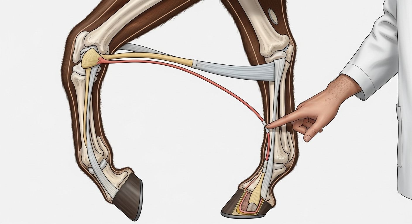
Zoom In: The Distal Limb—Where the Magic (and Most Injuries) Happen
Ah, the distal limb of the horse—a phrase that sounds like it belongs on a GCSE Biology paper, but honestly? It’s the *heart* of the anatomy of a horses leg. This zone runs from the fetlock joint down to the toe, and it’s where physics, anatomy, and fate collide every time hooves meet turf. Inside this compact marvel: the long and short pastern bones (P1 & P2), the coffin bone (P3, or pedal bone), the navicular bone (a tiny, grumpy-looking sesamoid with attitude), and the digital cushion—a spongy shock absorber that deserves a Royal Mail stamp in its honour. The distal limb of the horse carries ~60% of the horse’s body weight per stride at trot, and up to 200% during gallop. No pressure, luv. This part’s the reason why lameness exams start with “Where’s the heat? Where’s the pulse?”—because the distal limb of the horse is both miracle and vulnerability rolled into one sinewy package.
Meet the Stars: Bones, Tendons, and the Suspensory Apparatus in the anatomy of a horses leg
Let’s introduce the main cast of the anatomy of a horses leg, shall we? Top billing goes to the *suspensory ligament*—not your average ligament, mind you. This bad boy runs from the back of the knee/hock down, splits into two branches around the fetlock, and tucks under the sesamoid bones like a supportive hug. It’s the MVP of shock absorption. Then we’ve got the *superficial digital flexor tendon* (SDFT)—the spring in the stride—and the *deep digital flexor tendon* (DDFT), the stealthy one that slips *through* the sesamoid arch to attach to the coffin bone. And let’s not forget the *check ligaments*—proximal and distal—acting like backup singers keeping tension just right. All of these players work in concert to turn kinetic energy into elastic recoil. In short? The anatomy of a horses leg has more teamwork than a village cricket final on Bank Holiday Monday.
What Are the 3 F’s for Horses? (No, Not *That* Kind of F)
Right-o—before your mind wanders to the pub, let’s clarify: in equine circles, the 3 F’s for horses stand for *Feed, Farriery,* and *Fitness*. And guess what? All three orbit tightly around the anatomy of a horses leg. — Feed? A balanced diet (especially calcium:phosphorus ratio and trace minerals like copper & zinc) directly affects bone density and tendon integrity. — Farriery? A well-balanced trim or shoe isn’t just “neat”—it redistributes load across the hoof capsule, protecting the navicular apparatus and preventing strain up the chain. — Fitness? Gradual conditioning strengthens tendons *and* ligaments (yes, they adapt—but slower than muscle). Rush it? Hello, bowed tendon. Skimp on farriery? Say g’day to sheared heels. Ignore feed? Weak bone = stress fractures. So yeah—the 3 F’s for horses are the holy trinity keeping that anatomy of a horses leg ticking like Big Ben on a good day.
The Lower Limb Lingo: What’s the Lower Part of a Horse’s Leg Called, Anyway?
“What’s the lower part of a horse’s leg called?”—a question asked by every newbie at the livery yard while stroking a suspiciously patient cob. Technically? The distal limb. But in the field (quite literally), folk’ll say *below the knee/hock*, *from the fetlock down*, or—if they’re showing off—*the digit*. Yep, horses are digitigrade animals, meaning they walk on their *toes* (P3, to be precise), with the hoof acting like a reinforced toenail. The lower part of a horse’s leg includes:
- Fetlock joint (metacarpophalangeal)—the “ankle” that isn’t
- Pastern (P1 + P2)—sloping elegance with shock-absorbing flex
- Coffin joint (P2–P3 & navicular)—tiny, complex, vital
- Hoof capsule—horn, laminae, sole, frog: nature’s composite boot
Common Misconceptions: “Horses Have Knees” and Other Tall Tales
Let’s bust a few myths, shall we? First: that “knee” on the front leg? It’s actually a wrist. True story. Anatomically, it’s the *carpus*—equivalent to your wrist bones. The horse’s *real* knee is hidden up in the stifle (hindlimb), buried under muscle near the flank. And the hock? That’s their *ankle* (tarsus), not a knee. Confusing? Absolutely. But once you grasp it, the anatomy of a horses leg starts making sense:
| Horse Term | Anatomical Equivalent (Human) | Function in anatomy of a horses leg |
|---|---|---|
| “Knee” (front) | Wrist (carpus) | Shock absorption, fine-tuning stride |
| Hock (hind) | Ankle (tarsus) | Propulsion, leverage for push-off |
| Stifle | Knee (femorotibial joint) | Main hinge for hindlimb power |
| Fetlock | Knuckle (metacarpophalangeal) | Load-bearing flex point, spring loading |
Performance & Pathology: How the anatomy of a horses leg Holds Up (or Doesn’t) Under Pressure
Here’s a sobering stat: over 60% of lameness cases in performance horses originate in the distal limb—the very heart of the anatomy of a horses leg. Why? Because physics is *brutal*. At gallop, peak vertical forces on a single forelimb can hit **2.5x the horse’s body weight**. A 500 kg horse = ~1,250 kg of force *per stride* on one leg. Repeated, day after day. No wonder the SDFT is the most commonly injured structure in racehorses. But here’s the kicker: modern imaging (MRI, ultrasound, nuclear scintigraphy) shows that *microdamage accumulates silently*—weeks before heat, swelling, or lameness appear. The anatomy of a horses leg is resilient, yes—but not invincible. That’s why proactive care (hello again, 3 F’s for horses) matters more than flashy training regimes. Because no amount of lunging in circles fixes poor hoof-pastern alignment or chronic low-grade inflammation in the navicular bursa.
Putting It All Together: Why Understanding the anatomy of a horses leg Makes You a Better Rider, Owner, or Fan
Look—whether you’re mucking out at dawn in the Cotswolds, schooling dressage in Kent, or just sipping tea trackside at Ascot, knowing the anatomy of a horses leg changes how you *see* horses. You’ll spot a shortened stride *before* it becomes a limp. You’ll understand why that farrier spends 20 minutes on one foot. You’ll appreciate why rest days aren’t “lazy”—they’re *essential* for collagen realignment. And when someone says, “He’s just a bit stiff today,” you’ll *ask*: Where? When? On which lead? So here’s our little nudge: — Start at Riding London for the big picture, — Dive deeper into Learn for biomechanics made digestible, — And geek out properly with Hoof Anatomy of a Horse: Internal Structure—because the hoof *is* the foundation of the entire anatomy of a horses leg. Stay curious. Stay observant. And never underestimate the poetry in a horse’s step.
Frequently Asked Questions
What is the anatomy of the horse's legs?
The anatomy of the horse's legs is a highly specialised, non-muscular distal structure designed for speed and endurance. It comprises proximal muscles (above knee/hock), long tendons (SDFT, DDFT), the suspensory ligament, sesamoid bones, and the distal limb—featuring the fetlock, pasterns, coffin joint, and hoof. Crucially, there are no muscles below the knee or hock, making tendons and ligaments the primary movers and shock absorbers. This setup allows elastic energy storage and release—key to the horse’s famed economy of movement.
What is the distal limb of the horse?
The distal limb of the horse refers to the portion of the leg from the fetlock joint down to the toe—including the pastern bones (P1 & P2), coffin (pedal) bone (P3), navicular bone, digital cushion, and hoof capsule. Despite its small size, this region bears immense load (up to 200% of body weight during gallop) and houses critical structures for shock absorption and propulsion. Because it lacks muscle and relies on passive support, the distal limb of the horse is both biomechanically brilliant and highly susceptible to injury—hence its outsized role in lameness diagnosis.
What are the 3 F's for horses?
The 3 F's for horses are Feed, Farriery, and Fitness—three pillars of sound limb health directly tied to the anatomy of a horses leg. Proper Feed ensures bone mineralisation and tendon collagen integrity; skilled Farriery maintains hoof balance and reduces strain on joints/tendons; structured Fitness allows gradual adaptation of soft tissues to load. Neglect any one, and the delicate equilibrium of the distal limb collapses—often before clinical signs appear. Smart management means honouring all three, every single day.
What is the lower part of a horse's leg called?
The lower part of a horse's leg is anatomically termed the distal limb, though colloquially it’s described as “below the knee or hock.” It includes the fetlock (often mistaken for an ankle), pastern, coffin joint, and hoof. Functionally, it acts as a spring-loaded digit—horses walk on the tip of a single toe (P3), encased in the hoof. This region is devoid of muscle, relying entirely on passive tension from tendons and ligaments—making the lower part of a horse's leg both an engineering marvel and a high-maintenance masterpiece.
References
- https://www.vet.cornell.edu/departments-centers-and-institutes/baker-institute/equine-orthopaedics
- https://www.ncbi.nlm.nih.gov/pmc/articles/PMC7893452/
- https://www.aesculight.com/equine-lameness/anatomy-of-the-equine-limb/
- https://www.rvc.ac.uk/research/research-centres-and-facilities/structure-and-motion/equine-biomechanics