Hoof Anatomy of a Horse Internal Structure
- 1.
What is the anatomy of the horse hoof? Let’s crack it open like a Sunday roast—careful, now, it’s tender
- 2.
What is the most sensitive part of a horse hoof? Hint: it’s not where you’d tap with a hoof pick
- 3.
What are the things on horse hooves called? No, not “bits and bobs”—proper names, please
- 4.
What is the bottom of a horse's foot called? Sole? Frog? Or the whole lot?
- 5.
The hoof wall: nature’s carbon fibre, grown fresh weekly
- 6.
The frog: unsung hero, shock absorber, and circulatory pump—all in one squish
- 7.
The digital cushion: padding with purpose, like your nan’s favourite armchair
- 8.
Laminitis warning signs—because early action beats late panic
- 9.
Hoof balance: angles aren’t optional—they’re physics with consequences
- 10.
Where to go next? Keep the learning canter steady—no need to bolt
Table of Contents
hoof anatomy of a horse
What is the anatomy of the horse hoof? Let’s crack it open like a Sunday roast—careful, now, it’s tender
Ever stared at a horse’s foot and thought, “That’s just a fancy toenail, innit?” Oh, *dear*—you’ve been had smoother than a dodgy timeshare pitch in Skegness. The anatomy of the horse hoof isn’t just keratin and hope—it’s a biomechanical *sonnet*, written in collagen, blood, and centuries of wild gallops across moor and marsh. Outer wall? Tough as a docker’s handshake. Sole? Concave, protective, *alive*. Frog? That rubbery V? It’s a shock absorber, blood pump, *and* traction aid—like nature’s own Michelin star. Inside? Coffin bone, navicular, laminae interlocking like a zip on a Barbour. Miss one piece, and the whole verse stumbles. A proper hoof anatomy of a horse diagram doesn’t just label—it *sings* function, force, and fragility in one glance.
What is the most sensitive part of a horse hoof? Hint: it’s not where you’d tap with a hoof pick
Right—here’s the tea: the most sensitive part of a horse hoof isn’t the sole (though thin soles *hurt*), nor the wall (cracks sting, but slowly). It’s the laminae—those feathery, interlocking tissues that bind coffin bone to hoof wall. Think of them as Velcro forged by angels: strong, precise, *exquisitely* nerve-rich. Inflame them (hello, laminitis), and your horse’s in agony—not from “ouch, stone”, but from *structural betrayal*. The sensitive laminae (attached to P3) are packed with nociceptors—more per cm² than your fingertips. That’s why a laminitic horse stands like he’s on hot coals, rocking back, eyes wide. Respect the hoof anatomy of a horse, and you’ll spot early signs—heat, digital pulse, reluctance to turn—before the crisis hits.
What are the things on horse hooves called? No, not “bits and bobs”—proper names, please
Down the yard, you’ll hear: “frog’s gone mushy”, “white line’s blown”, “bars need trimming”. But what *are* these “things on horse hooves called”? Let’s roll call—properly:
- Hoof wall — the tough outer shell (toe, quarters, heels)
- Sole — the concave underside (not weight-bearing, *protective*)
- Frog — central V, elastic, grooved
- Bars — inward folds of wall, from heels to mid-frog
- White line — the *junction* (not white!—yellowish), where wall meets sole
- Periople — the waxy “cuticle” at the coronet
- Heel bulbs — the soft, fleshy pads behind the frog
Get these wrong, and you’ll sound like someone ordering a “tea with milk” in a Glasgow pub—technically correct, but *missing the nuance*. A crisp hoof anatomy of a horse chart labels every one—often colour-coded, because yes, the white line *is* a bit beige. Go figure.
What is the bottom of a horse's foot called? Sole? Frog? Or the whole lot?
Trick question! The bottom of a horse's foot isn’t *one* thing—it’s a *landscape*. Technically? The entire ground-facing surface is the sole region, but it’s a mosaic: central frog, surrounding sole (calloused, not flat), flanking bars, and the critical white line hugging the inner wall. In farrier terms, “the sole” = the keratin plate *between* frog and wall—not the whole underside. Confuse them, and you’ll pare too deep, expose the coffin bone, and spend £300 on remedial pads and tears. A top-tier hoof anatomy of a horse illustration shades these zones distinctly—because “bottom” is *geography*, not gossip.
The hoof wall: nature’s carbon fibre, grown fresh weekly
Ah, the hoof wall—laminated keratin, laid down in tubules like terraced cottages on a Welsh hillside. It grows ~1 cm/month (faster in summer, slower in winter—just like your will to mow the lawn). It’s *not* dead tissue—it’s *alive* at the coronet, nourished by blood, responding to load like memory foam with ambition. Cracks? Flares? Seedy toe? All red flags—often from imbalance, poor diet (low biotin, high sugar), or infrequent trimming (£45–£75 every 6–8 weeks, luv—worth every penny). In any decent hoof anatomy of a horse cross-section, the wall’s the bold outer rim—thick, layered, *resilient*. But resilience isn’t infinite. Respect the growth, or you’ll pay in lameness later.
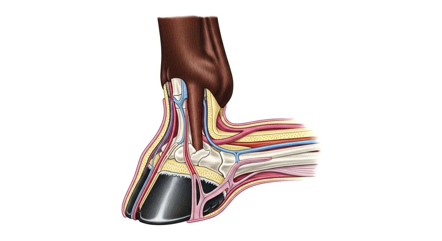
The frog: unsung hero, shock absorber, and circulatory pump—all in one squish
Right in the middle—rubbery, grooved, often underestimated—is the frog. Press it? It yields, then *recoils*. That’s the hoof pump mechanism in action: compressing the frog squishes blood from the digital cushion up the leg—like giving the circulatory system a gentle nudge with a teaspoon. No frog contact? Poor circulation. No elasticity? Stiff, cold feet. Thrush? A foul-smelling surrender to damp and neglect. Healthy frogs are wide, firm, and split cleanly down the middle—like a proper Sunday roast joint. In a hoof anatomy of a horse diagram, it’s usually shaded darker red—because functionally, it’s *the heart* of the foot.
The digital cushion: padding with purpose, like your nan’s favourite armchair
Tucked above the frog—soft, fibro-fatty, criminally overlooked—is the digital cushion. Think of it as the hoof’s airbag. At impact, it spreads, pushes the heels *outward*, and helps the whole capsule expand. Foals? Mostly fat. Adults? Fibrous tissue takes over—*especially* in barefoot horses with proper ground contact. Shod too long? Cushion atrophies. Result? Poor shock absorption, navicular strain, and heels that cave inward like soggy biscuits. A good hoof anatomy of a horse schematic shows it as a pale wedge—often labelled “DC”—because without it, the hoof’s like a crisp packet with no crunch: all form, no function.
Laminitis warning signs—because early action beats late panic
Laminitis isn’t “just hoof pain”—it’s a *systemic betrayal*. Here’s what to watch for (BEVA 2024 UK field data):
| Sign | Early (0–24h) | Acute (24–72h) | Action Window |
|---|---|---|---|
| Hoof heat | +2–3°C above ambient | +5°C+, radiating | Golden hour: vet NOW |
| Digital pulse | Faintly bounding | Throbbing, “wire-like” | Check daily in at-risk horses |
| Stance | Reluctant to turn | “Sawhorse” posture, rocked back | Box rest + deep bedding |
| Gait | Shortened stride | Extreme heel-first landing | No forced walking! |
“It’s not the hoof that fails—it’s the system around it,” as the old farrier used to mutter, stirring his tea. A detailed hoof anatomy of a horse doesn’t just show parts—it maps *vulnerability*.
Hoof balance: angles aren’t optional—they’re physics with consequences
Ideal front hoof angles? Hoof wall (toe): 50–55° Pastern: 45–55° Hoof-pastern axis: *straight line*—no “broken back” or “broken forward” Deviate? Short, upright hooves = more concussion = ringbone risk. Long, low heels = strain on DDFT = navicular syndrome. Even the *width* matters: heels should match frog width—not pinched, not flared. A balanced hoof isn’t “pretty”—it’s *sustainable*. And any proper hoof anatomy of a horse guide overlays angles on photos—because numbers without visuals? Like GPS without signal.
Where to go next? Keep the learning canter steady—no need to bolt
Had your fill? Nah—you’ve just warmed up the kettle. For the full lowdown on equine biomechanics—from poll to pastern—start at the hub: Riding London. Fancy structured learning? Our Learn section’s got vet-reviewed guides, trimming videos, and lameness checklists. And if you’re *really* itching to geek out on the nitty-gritty, don’t miss our ultra-detailed companion piece—anatomy of hoof horse: detailed sole diagram—with layer-by-layer dissection, pathology markers, and farrier notes. Because knowledge? That’s the best bit of kit you’ll ever buy—and it doesn’t need re-greasing.
Frequently Asked Questions
What is the anatomy of the horse hoof?
The anatomy of the horse hoof comprises external structures—the hoof wall (toe, quarters, heels), sole, frog, bars, and white line—and internal components: the coffin bone (P3), navicular bone, digital cushion, laminae (sensitive + insensitive), and vascular networks. It functions as a dynamic shock absorber, circulatory pump, and weight-bearing platform. A detailed hoof anatomy of a horse diagram integrates all layers to show structural and functional interdependence.
What is the most sensitive part of a horse hoof?
The most sensitive part of a horse hoof is the sensitive laminae—the vascular, nerve-rich tissue attaching the coffin bone to the inner hoof wall. These contain a high density of nociceptors, making them acutely responsive to inflammation (e.g., laminitis). Even minor separation causes significant pain. Hence, understanding hoof anatomy of a horse prioritises lamellar health as foundational to soundness and welfare.
What are the things on horse hooves called?
The key things on horse hooves called include: hoof wall (outer protective layer), sole (concave underside), frog (central V-shaped pad), bars (wall folds along frog margins), white line (junction between wall and sole), periople (coronary sealant), and heel bulbs (posterior soft tissue). Correct terminology is vital for farrier communication and clinical accuracy—each feature has biomechanical significance in the hoof anatomy of a horse.
What is the bottom of a horse's foot called?
The bottom of a horse's foot is collectively termed the *solar surface* or *ground surface*, composed of the sole, frog, bars, and white line. While “sole” is often used colloquially for the whole underside, technically it refers only to the keratinised plate *between* the frog and inner wall. Precision matters: misidentifying regions can lead to improper trimming or missed pathology. A comprehensive hoof anatomy of a horse guide distinguishes these zones clearly for clinical application.
References
- https://www.ncbi.nlm.nih.gov/pmc/articles/PMC6383295
- https://www.bevalibrary.org/horse-hoof-anatomy
- https://vet.osu.edu/vmc/companion/our-services/horse-keeping-hint/horse-hoof-anatomy