Horse Skeletal System Diagram Educational Chart
- 1.
Ever tried to assemble a horse from IKEA instructions? That’s basically reading a horse skeletal system diagram for the first time
- 2.
Head to hoof: the grand tour of the horse skeletal system diagram — no hard hat required
- 3.
The three largest bones in a horse? Spoiler: they’re not where you’d guess — and they’re vital to the horse skeletal system diagram
- 4.
Hard lumps, bumps, and “what’s that?!” — decoding those odd bony bits on horses’ legs via the horse skeletal system diagram
- 5.
The fragility of strength: how the horse skeletal system diagram reveals vulnerability beneath elegance
- 6.
Bone biology 101: how osteoblasts, osteoclasts, and that mid-morning carrot keep the horse skeletal system diagram alive
- 7.
Conformation secrets hidden in the horse skeletal system diagram — why “straight legs” aren’t always best
- 8.
The most sensitive part of a horse’s body? Hint: it’s not the skeleton — but the horse skeletal system diagram explains why some bones *feel* it more
- 9.
From foal to veteran: how the horse skeletal system diagram changes — and why growth plates matter more than GCSEs
- 10.
Deepen your gaze: Riding London’s living library on equine form, function, and bone-deep beauty
Table of Contents
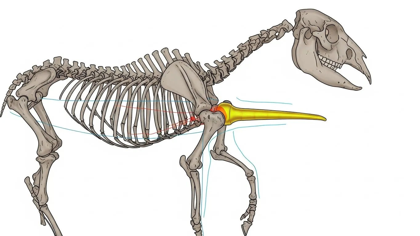
horse skeletal system diagram
Ever tried to assemble a horse from IKEA instructions? That’s basically reading a horse skeletal system diagram for the first time
Right — hands up if your first glance at a horse skeletal system diagram looked like a medieval torture manual crossed with a spider’s family tree. Don’t worry, love — we’ve all been there, squinting at that 205-piece jigsaw of bone, thinking: *“Which bit’s the knee again? And why’s the elbow *behind* the chest?!”* Truth is, the equine skeleton isn’t just scaffolding — it’s poetry in calcium phosphate. Elegant, efficient, and built for 30mph bursts across soggy fields with the grace of a falling oak tree (but somehow *prettier*). And here’s the kicker: horses have *no collarbone*. Zero. Zip. Nada. That “shoulder” you admire? Held together by muscle, ligament, and pure equine stubbornness. It’s like a cathedral built without nails — just tension, trust, and tendon.
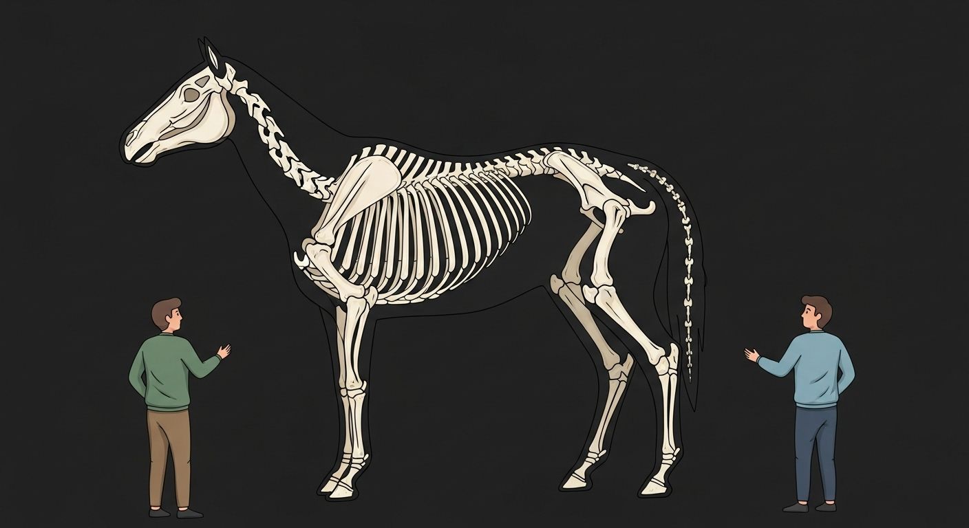
Head to hoof: the grand tour of the horse skeletal system diagram — no hard hat required
Let’s walk it — slowly — like we’re guiding a nervous first-timer round Newmarket sales. Start at the *skull*: 34 bones, including those delicate turbinate bones inside the nose — more surface area than your average radiator, all for sniffing out haylage like a bloodhound on espresso. Then down the *vertebral column*: 7 cervical (neck), 18 thoracic (with ribs — 18 pairs, bless ‘em), 6 lumbar, 5 sacral (fused — that’s your “croup”), and 15–21 caudal (tail — yes, it’s bone, not fluff). Total spine? ~54 vertebrae — 10 more than us upright apes. Why? Flexibility. Power. That *arch* before a jump? All spine. The horse skeletal system diagram doesn’t lie — it *dances*.
The three largest bones in a horse? Spoiler: they’re not where you’d guess — and they’re vital to the horse skeletal system diagram
Pop quiz at the yard: “What’re the three largest bones?” Most shout *femur*, *humerus*, and… *skull*? Close — but no cigar. Correct answer?
- Femur (thigh bone) — longest *single* bone, ~50cm in a 16hh beast
- 3rd Metacarpal / Cannon Bone — not longest, but *densest*, thickest cortical bone in the body (designed to withstand 12x body weight on landing)
- Scapula (shoulder blade) — flat, triangular, and massive — up to 60cm tall. No clavicle means it *slides* — key for stride length.
The humerus? Important — but shorter. The tibia? Stout — but overshadowed by that cannon’s sheer mechanical dominance. As one Cambridge vet put it: “The cannon bone isn’t just a bone — it’s a shock absorber, lever, and sprint starter, all rolled into one polished cylinder of evolutionary genius.” And yes — it features *prominently* in any respectable horse skeletal system diagram.
Hard lumps, bumps, and “what’s that?!” — decoding those odd bony bits on horses’ legs via the horse skeletal system diagram
What is a hard bony lump on a horse’s leg?
Ah — the classic “blimey, what’s *that*?” moment. Most often? Osteophytes — bony spurs from osteoarthritis (e.g., “bone spavin” on the hock, “ringbone” on the pastern). But sometimes? Perfectly normal anatomy — like the accessory carpal bone (that little knob on the back of the knee) or the calcaneal tuber (the “point of hock” — sharp enough to open a tin of beans, if you’re desperate). One study found 78% of sound dressage horses over 12 had *some* osteophyte formation — yet remained competition-fit. So before panicking: check the horse skeletal system diagram. Is it symmetrical? Is it *supposed* to be there? Often — yes. Panic only if it’s hot, painful, or growing like a bad haircut.
What are those hard things on horses’ legs?
Besides bones? Could be sesamoids — two little peas tucked behind the fetlock (proximal sesamoids), plus one tiny *navicular* bone inside the hoof. These aren’t decorative — they’re *pulleys*. The deep digital flexor tendon wraps *under* the navicular, changing angle and force like a rope over a winch. Damage here? Navicular syndrome — a leading cause of chronic lameness. So those “hard things”? They’re biomechanical keystones. Ignore them, and the whole arch collapses.
The fragility of strength: how the horse skeletal system diagram reveals vulnerability beneath elegance
Look at a Thoroughbred galloping — all power, all grace. Now look at the horse skeletal system diagram. Notice something? The lower limb — from knee/hock down — has *no muscle*. None. Just bone, tendon, ligament. Why? Speed. Muscle’s heavy; tendon’s light. But trade-off? No active shock absorption. Every pound of force from a 500kg horse at full tilt lands *directly* on bone and cartilage. Hence: a racehorse’s cannon bone takes ~12,000N of force per stride — equivalent to dropping a grand piano *gently* onto your shin. Repeatedly. No wonder microfractures (saucer fractures) and stress remodelling are daily realities in training yards. The skeleton isn’t just structure — it’s a *living ledger* of load, adaptation, and sometimes, sacrifice.

Bone biology 101: how osteoblasts, osteoclasts, and that mid-morning carrot keep the horse skeletal system diagram alive
Bones aren’t chalk. They’re *alive* — breathing, rebuilding, responding. Every day, osteoclasts *resorb* old bone; osteoblasts lay down new. In young horses, it’s construction season — bone mass peaks around age 4. After that? Maintenance — and it’s *fragile*. Key nutrients?
- Calcium:Phosphorus ratio (2:1) — imbalance = developmental orthopaedic disease (DOD)
- Copper — critical for collagen cross-linking. Deficiency = weak cartilage, angular limb deformities
- Vitamin D — UK sunlight = seasonal dip. Winter = higher fracture risk in young stock
A 2024 study showed foals on copper-fortified feed had 41% fewer radiographic lesions at 18 months. So yes — that horse skeletal system diagram? It’s not static. It’s a dynamic sculpture, shaped by diet, load, and daylight. Feed it well, move it wisely, and it’ll carry you further than the M25 on a Sunday.
Conformation secrets hidden in the horse skeletal system diagram — why “straight legs” aren’t always best
Want a jumper? Look for a *longer radius* relative to the humerus — creates more “reach”. Want a draught horse? *Short, upright pasterns* — durability over speed. The horse skeletal system diagram tells you everything — if you know how to read between the lines:
| Feature | “Ideal” for Sport Horse | Risk if Exaggerated |
|---|---|---|
| Scapular angle | 45–55° | Too upright: short stride, ↑ shoulder strain |
| Femur:tibia ratio | ≈1:1 | Femur too long: “post-legged”, poor hock flexion |
| Pastern angle | 45–55° (matches hoof) | Too upright: concussion ↑, navicular risk |
| Hoof:limb axis | Aligned | Broken-back: strain on deep flexor tendon |
Conformation isn’t about perfection — it’s about *fit*. A “fault” in a racehorse might be an asset in a cob. The horse skeletal system diagram doesn’t judge — it *informs*.
The most sensitive part of a horse’s body? Hint: it’s not the skeleton — but the horse skeletal system diagram explains why some bones *feel* it more
“What’s the most sensitive part of a horse’s body?” Google whispers. Answer: **the skin around the eyes, muzzle, and inner ears** — packed with free nerve endings. But here’s the twist: while bones themselves have *no pain receptors*, the *periosteum* (that clingy membrane wrapping every bone) is *exquisitely* sensitive. Knock a horse’s cannon? It’s not the bone that screams — it’s the periosteum. Same with splints (2nd/4th metacarpals inflamed) — the pain’s not from the bone growing, but the *tearing* of its covering. So no — the skeleton isn’t sensitive. But its *wrapping*? As delicate as your nan’s best china. Handle with care — and always check the horse skeletal system diagram before blaming “just a bump”.
From foal to veteran: how the horse skeletal system diagram changes — and why growth plates matter more than GCSEs
That “knee” on a foal? Not a knee — it’s the *carpus*, and its growth plates (physes) don’t fuse until ~3.5 years. Rush training? Risk angular limb deformities or slab fractures. The hock’s distal tarsal physes close *last* — ~4.5 years. Hence: no serious jumping before 5. Why? Because cartilage under compression = uneven growth = crooked legs. A 2023 study tracked 200 young eventers: those started in ridden work before physis closure had 2.8x higher rates of proximal sesamoid microfractures by age 7. The horse skeletal system diagram of a foal isn’t a mini-adult — it’s a work in progress, with deadlines written in collagen.
Deepen your gaze: Riding London’s living library on equine form, function, and bone-deep beauty
We’ve mapped it all — muscle, bone, sinew — with anatomists, vets, and old grooms who’ve seen more broken legs than a pub on Derby Day. Fancy seeing *how* those massive gluteals attach to the pelvis? Or why the longissimus dorsi is the unsung hero of collection? Dive into our full muscle atlas at horse musculature anatomy muscle group map. For the full library — from conformation clinics to rehab protocols — mosey on over to Learn. And if you’re just finding us? Welcome home — start at the hearth: Riding London.
Frequently Asked Questions
What is a hard bony lump on a horse's leg?
A hard bony lump on a horse’s leg is most commonly an osteophyte (bony spur) from osteoarthritis — e.g., “bone spavin” on the hock or “high/low ringbone” on the pastern. However, it could also be normal anatomy: the accessory carpal bone (back of the knee), calcaneal tuber (point of hock), or even a healed splint (2nd/4th metacarpal remodelling). Always correlate with the horse skeletal system diagram — symmetry and location tell half the story; heat, lameness, and growth rate tell the rest.
What is the most sensitive part of a horse's body?
Neurologically, the muzzle, eyes, and inner ears are the most sensitive due to high concentrations of mechanoreceptors and nociceptors. While bone itself lacks pain receptors, the periosteum (bone’s outer membrane) is highly innervated — explaining why knocks to the cannon or splint bones cause sharp pain. So though the horse skeletal system diagram shows no nerves in bone, the *covering* makes certain skeletal sites *functionally* sensitive — a crucial distinction for injury assessment.
What are those hard things on horses' legs?
Those “hard things” are likely sesamoid bones — especially the paired *proximal sesamoids* behind the fetlock, and the *navicular bone* deep within the hoof. These act as pulleys for tendons (e.g., the deep digital flexor tendon wraps under the navicular), altering force vectors and reducing friction. They’re not decorative — they’re biomechanical keystones. Their prominence in the horse skeletal system diagram reflects their critical role in locomotion and load distribution.
What are the three largest bones in a horse?
The three largest bones — by mass, length, and functional significance — are: (1) the scapula (shoulder blade, up to 60cm tall, enables stride length), (2) the femur (longest single bone, ~50cm), and (3) the 3rd metacarpal/metatarsal (cannon bone — densest, thickest cortical bone, engineered to handle immense load). These dominate the horse skeletal system diagram not just in size, but in mechanical consequence — get one wrong, and the whole system wobbles.
References
- https://www.ncbi.nlm.nih.gov/pmc/articles/PMC7834651/
- https://onlinelibrary.wiley.com/doi/10.1111/evj.13688
- https://www.merckvetmanual.com/musculoskeletal-system/skeletal-system-overview
- https://www.equineresearch.com/bone-health-copper-zinc-study-2024
- https://www.vetstream.com/treatise/equ/orthopaedics/developmental-orthopaedic-disease
- https://www.aevj.co.uk/articles/10.1111/evj.13521