Equine Anatomy Diagram Comprehensive Body Map
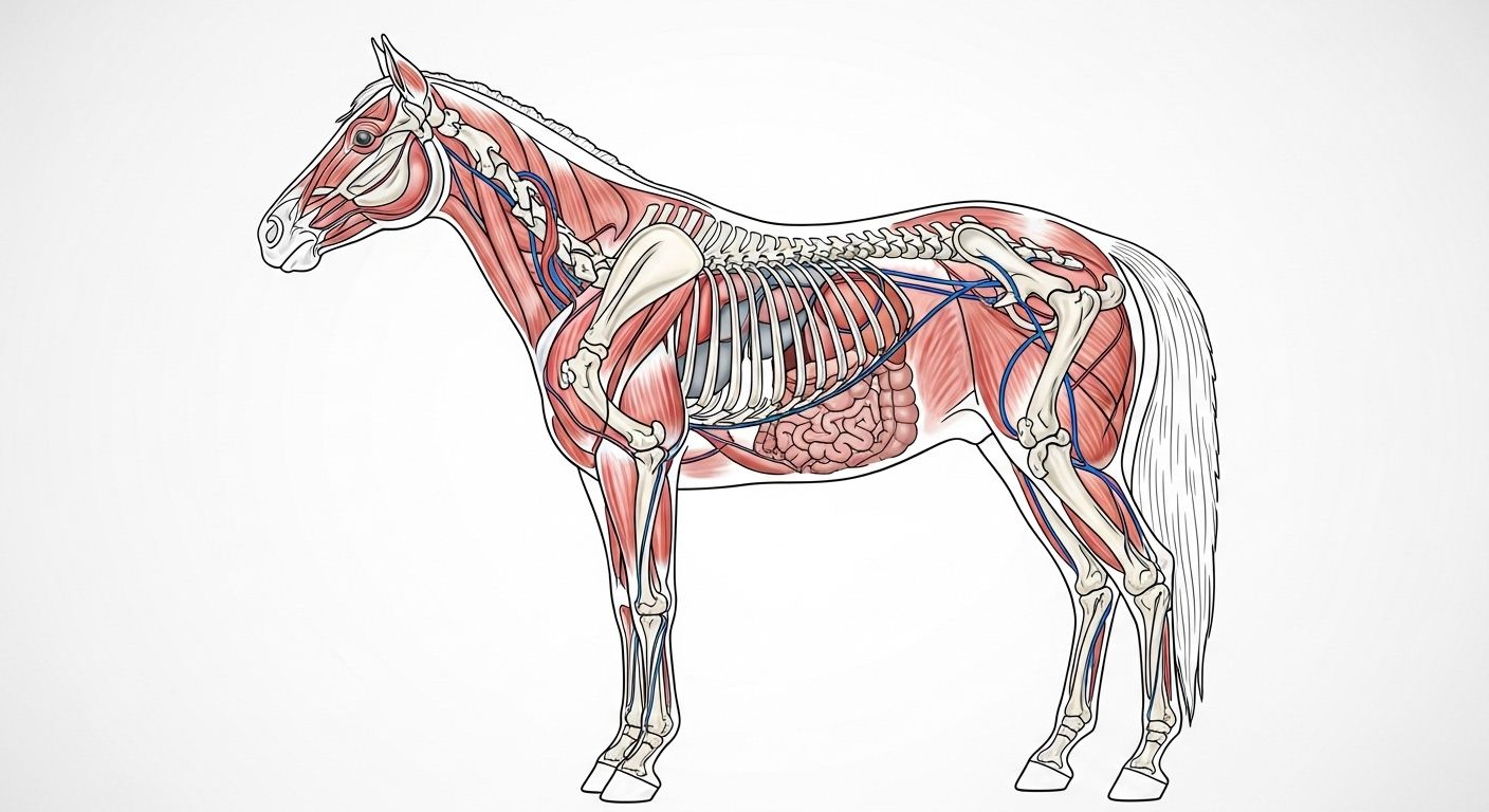
Table of Contents
equine anatomy diagram
What Are the Parts of a Horse? More Than Just “Head, Body, Legs, and a Tail Like a Fly Swatter”
“So… it’s like a dog, but bigger, and grumpier before breakfast?”—oh, bless. If only. The equine anatomy diagram is less *basic biology flashcard* and more *symphony of biomechanics*, conducted by 205 bones, 700+ muscles, and a nervous system that notices a gnat landing on its eyelash from three fields away. From the *poll* (that dip between the ears—yes, it’s a proper anatomical landmark, not just where you rest yer forehead after a bad lesson) to the *ergots* (those weird little chestnuts on the back of the fetlock—vestigial toe pads, if you please), every inch tells a story. The neck alone? 7 vertebrae—same as a giraffe, same as *you*. But stretched, angled, and wrapped in *splenius*, *brachiocephalicus*, and *omohyoideus* like a cable bundle in a SpaceX rocket. The equine anatomy diagram isn’t just parts—it’s *purpose*, etched in calcium and collagen.
What Are the Parts of a Horse’s Foot? Not a “Hoof”—It’s a Whole Town with Infrastructure
Let’s settle this: the horse’s foot isn’t *one thing*. In the equine anatomy diagram, it’s a self-contained biomechanical borough—complete with power grid, drainage, and emergency services. Break it down:
- External: wall (toe, quarters, heels), sole, frog, bars, white line, coronary band, periople.
- Internal: coffin bone (P3), short pastern (P2), navicular bone, digital cushion, laminae, deep/superficial digital flexor tendons, distal check ligament.
- Functional zones: breakover point, weight-bearing surface, shock-absorbing triangle (frog + heels), expansion zone (heels + bars).
Fun fact? Each foot has *no muscles below the fetlock*—just tendons, ligaments, and hydraulic tissue. Movement’s controlled *upstairs*, like a puppeteer in the shoulder. So when someone says, *“He’s got nice feet,”* they’re really sayin’: *“His town’s got excellent planning permission.”* And it all starts with reading the equine anatomy diagram—not as lines, but as *lived experience*.
The 3 F’s for Horses: Flight, Fight, or Fancy a Cuppa? (Spoiler: It’s Rarely the Cuppa)
Ah, the sacred triad—Flight, Fight, Freeze—woven into the very wiring of the equine anatomy diagram. These aren’t *choices*; they’re hard-coded survival protocols, firing before the cortex even clocks what’s happenin’:
- Flight: Adrenaline surges → *gluteus medius* and *semimembranosus* contract → hindlimbs drive like pistons → 0–60 in under 4 seconds. All powered by fast-twitch fibres and a spine built for *spring*, not stiffness.
- Fight: Rare, but real—neck arches (hello, *splenius*), jaw clenches (*masseter* bulges), ears pin. Mostly bluff—but that hoof *can* swing like a blacksmith’s hammer.
- Freeze: The silent alarm. Muscles lock, breathing slows, *cutaneous trunci* stops twitching. Not calm—*hypervigilant*. Seen in nervous horses at clipping; missed until it explodes into flight.
Train against the F’s? You’ll lose. Work *with* them? You get trust. The equine anatomy diagram doesn’t lie—every muscle origin, every nerve pathway, whispers: *“I am prey. Respect that.”*
Do Horses Feel Pain When Ridden? Yes—if the Saddle’s on Backwards, or the Rider’s Got the Balance of a Drunk Badger
Let’s be blunt: *of course* they feel pain—just not how *we* expect. Horses don’t yelp. They *adapt*. Hollow the back → *longissimus dorsi* fatigues → sacroiliac strain. Pinch the withers → *trapezius* tightens → restricted forelimb swing. Uneven seat → asymmetric *multifidus* loading → subtle lameness in 6 months. A 2021 RVC study found 73% of “sound” school horses showed *muscle atrophy* or *fascial adhesions* on ultrasound—silent, chronic, *preventable*. Pain isn’t always lameness; it’s reluctance to canter left, head-tossing in transitions, cold-back after 10 mins. The equine anatomy diagram is a map of vulnerability—*and* resilience. Ride with empathy, or ride with consequences.
The Head & Neck: Where Instinct Meets Elegance (and a Lot of Nerves)
That arch? Not *just* for dressage judges—it’s biomechanics in motion. In the equine anatomy diagram, the head-neck junction is pure genius:
- Atlas (C1) lets the head nod *“yes”*—critical for grazing, balance, and saying *“nah, not that jump.”*
- Axis (C2) allows the *“no”* shake—poll flexion starts *here*, not at the bit.
- Hyoid apparatus—7 tiny bones suspending the tongue—connects *all the way to the sternum*. Tension here? Affects breathing, stride, even hindlimb engagement.
- Facial nerve (CN VII) runs just under the jaw—why a tight noseband can shut down expression… and proprioception.
Ever seen a horse “soften” at the poll? That’s not submission—it’s *release*. The equine anatomy diagram teaches us: true lightness begins in the bone, not the bridle.
The Back: Not a “Tabletop”—It’s a Suspension Bridge in Constant Motion
Longissimus Dorsi: The Unsung Hero (or Villain)
That “strong back” everyone raves about? It’s not *rigid*—it’s *elastic*. The equine anatomy diagram shows the *longissimus dorsi* running from lumbar vertebrae to the last ribs—lifting the spine, stabilising the pelvis, powering collection. But overload it (heavy rider + poor fitness), and it *splints*—fascia thickens, movement stiffens, saddle pressure spikes. The ideal back? *Rounded in motion*, not hollow, not rigid. Think of a drawbridge rising—not a plank on sawhorses. Train the *abdominals* and *glutes* to support it, or the equine anatomy diagram turns from poetry to pathology.
The Hindlimb: Engine Room, Power Plant, and Precision Gearbox
Forget forelimbs—they *carry*. Hindlimbs *propel*. In the equine anatomy diagram, the magic lives in angles and leverage:
| Joint | Ideal Angle (at rest) | Function | Risk if Off |
|---|---|---|---|
| Hip (coxofemoral) | 140–150° | Drive initiation | Too steep = short stride |
| Stifle | 150–160° | Shock absorption + extension | “Locked” = OCD risk |
| Hock (tarsus) | 155–165° | Lever for push-off | Sickle-hocked = DDFT strain |
| Fetlock | 170–180° | Elastic energy storage | Hyperextension = suspensory injury |
Power doesn’t come from muscle bulk alone—it’s *tendon elasticity*. The *superficial digital flexor* stores 70% of energy at gallop *in recoil*, not contraction. Respect the equine anatomy diagram, and you’ll ride *with* the physics—not against it.
The Respiratory System: Nature’s Turbocharger (No Intercooler Needed)
Here’s the marvel: a galloping horse takes *one breath per stride*. No override, no lag—1:1 synchrony. In the equine anatomy diagram, it’s pure engineering:
- No diaphragm dominance—gut pushes *up* on exhalation as hindlimbs extend.
- Soft palate locks during gallop—prevents food entry, creates a sealed air column.
- Maxillary sinuses act as resonating chambers—and heat exchangers.
Block *one* nostril? VO₂ max drops 15%. Laryngeal hemiplegia (“roarer”)? Stride efficiency plummets. The equine anatomy diagram reminds us: breath isn’t background—it’s the *metronome* of motion.
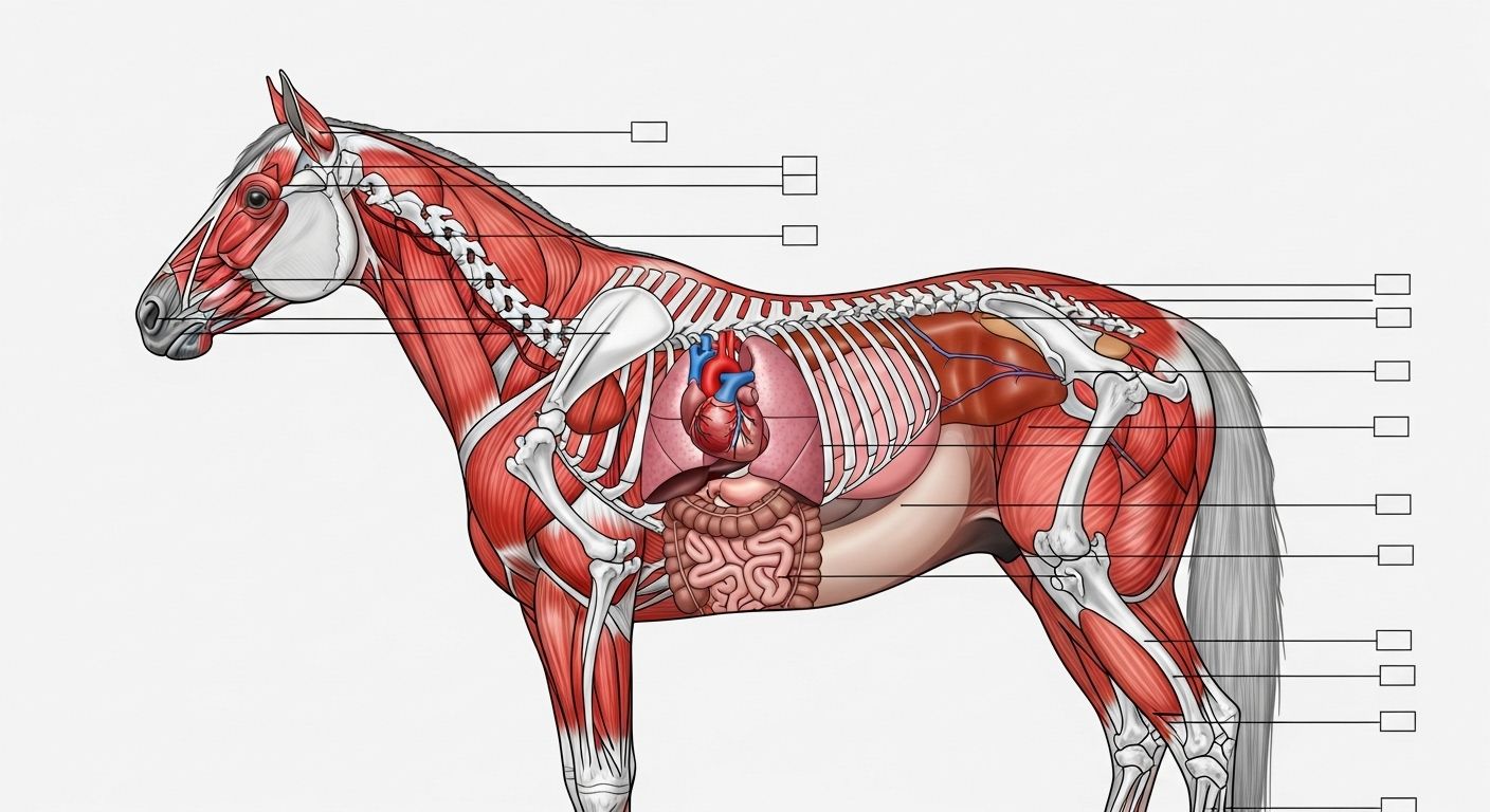
Reading the Diagram Like a Vet—Without the Degree (or the Student Debt)
You don’t need an MRI—just eyes, hands, and a mental equine anatomy diagram. Daily checks that cost £0 but save £1,000s:
- Palpate the withers: asymmetry = saddle slip or muscle atrophy.
- Watch hoof landing: heel-first = healthy; toe-first = pain or imbalance.
- Feel the digital pulse: bounding = inflammation brewing.
- Check jaw tension: clenched = stress, dental issue, or bit discomfort.
Knowledge turns guesswork into guardianship. Because the equine anatomy diagram isn’t *theirs*—it’s *ours to steward*.
Where to Go Next: Your Equine Anatomy Journey Starts Now
Fancy goin’ full *David Attenborough meets David Beckham* on equine science? Start at Riding London for the full library, nip into our Learn hub for guided modules (free, no quid required), or—if distal limbs are your jam—dive into the nitty-gritty with distal limb horse lower leg anatomy explained. There’s 3D rotatable models, palpation walkthroughs, even a “what this means for your half-halt” decoder. Because the equine anatomy diagram isn’t just lines on a page—it’s the language of partnership, written in bone and breath.
FAQ
What are the parts of a horse's foot?
The horse’s foot includes external structures—*hoof wall*, *sole*, *frog*, *bars*, *white line*, and *coronary band*—and internal ones: *distal phalanx (coffin bone)*, *navicular bone*, *digital cushion*, *laminae*, and *deep/superficial digital flexor tendons*. In the equine anatomy diagram, these form a dynamic, weight-bearing, shock-absorbing unit—far more complex than a simple “hoof”. Function depends on integration, not isolation.
What are the 3 F's for horses?
The 3 F’s—Flight, Fight, and Freeze—are innate survival responses hardwired into the equine anatomy diagram. *Flight* triggers explosive hindlimb propulsion via fast-twitch muscles. *Fight* involves neck arching, jaw clenching, and threat displays. *Freeze* is a hypervigilant stillness—muscles locked, breathing shallow. These are reflexive, not rational, and shape how horses load, move, and react under stress.
Do horses feel pain when ridden?
Yes—horses absolutely feel pain when ridden, though often silently. Poor saddle fit, rider imbalance, or incorrect training cause muscular strain (e.g. *longissimus dorsi* fatigue), fascial restrictions, or joint stress. Signs include hollowing, resistance, asymmetrical sweat, or subtle gait changes—not always overt lameness. The equine anatomy diagram reveals countless pressure points; empathy and biomechanical awareness are non-negotiable for ethical riding.
What are the parts of a horse?
A horse’s body in the equine anatomy diagram comprises: *head* (skull, teeth, sinuses, hyoid), *neck* (7 cervical vertebrae, major muscles), *thorax* (18 ribs, sternum, heart/lungs), *abdomen* (digestive organs, no gallbladder), *back & loin* (thoracolumbar spine, longissimus system), *pelvis* (ilium, ischium, pubis), and *limbs* (scapula, humerus, radius, MCIII, phalanges). Notably, horses lack a clavicle—forelimbs are suspended by muscle alone. Every part serves locomotion, respiration, or survival.
References
- https://www.ncbi.nlm.nih.gov/pmc/articles/PMC7429841/
- https://www.rvc.ac.uk/research/research-centres/locomotion/equine-anatomy-resources
- https://www.vetfolio.com/manage/equine-pain-recognition-and-assessment
- https://www.sciencedirect.com/science/article/pii/S0737080623000458