Distal Limb Anatomy Horse Foot to Knee Details
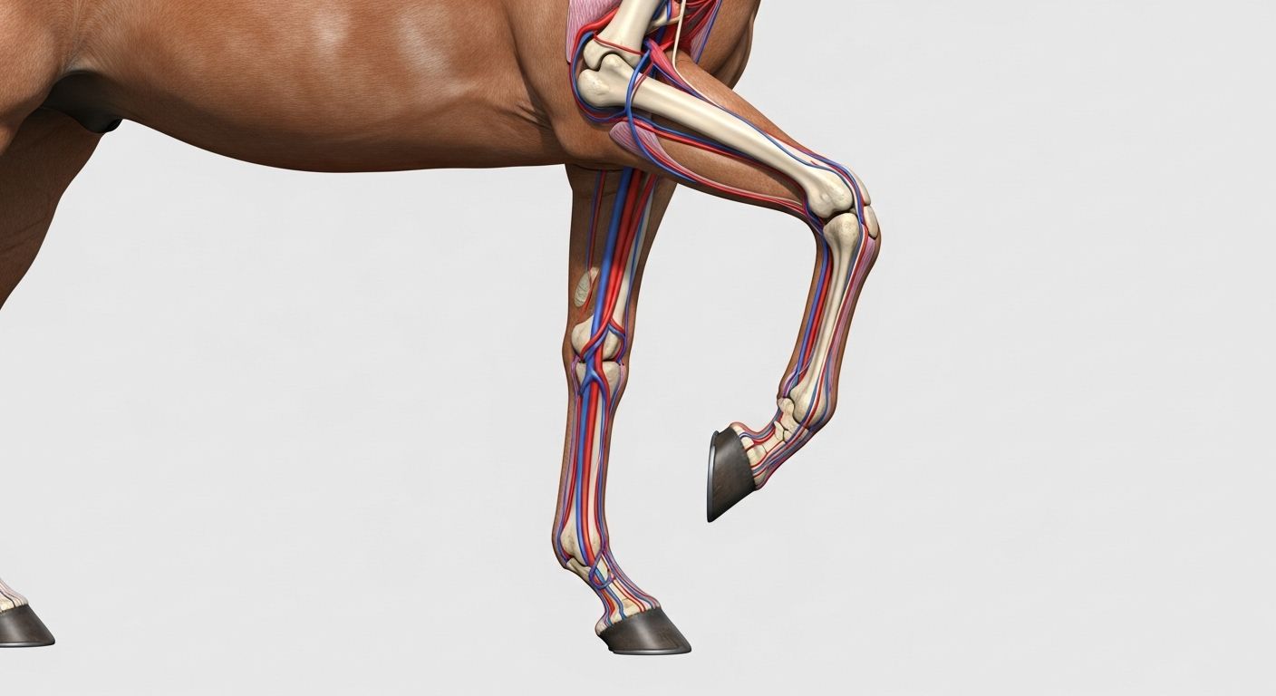
Table of Contents
distal limb anatomy horse
What Is the Distal Limb of the Horse? Not Just “Below the Knee”—It’s a Masterclass in Evolutionary Engineering
“So… it’s like a human hand, but backwards and covered in keratin?”—mate, if only biology were that tidy. The distal limb anatomy horse is *not* a hand. It’s **one toe**, encased in a hydraulic capsule, suspended by rope-like tendons, and fine-tuned over 55 million years to do *one thing* brilliantly: run like the wind *without* knackerin’ itself. From the carpus (“knee” in forelimb) or tarsus (“hock” in hind) down—it’s all *distal*. No muscles. No fat. Just bone, tendon, ligament, and the toughest biological composite on earth: hoof. Get this bit wrong, and the whole symphony falters. Nail it? You’re watchin’ biomechanics poetry in motion.
Proximal vs Distal on a Horse? Think Tube Map, Not Geography
Right—before we dive in, let’s clear the fog. “Proximal” = *closer to the core* (like Oxford Circus on the Central Line). “Distal” = *farther out* (hello, Epping). In the distal limb anatomy horse, that means:
- Proximal forelimb: shoulder → humerus → elbow.
- Distal forelimb: “knee” (carpus) → cannon (MCIII) → pasterns (P1, P2) → coffin bone (P3).
- Proximal hindlimb: pelvis → femur → stifle.
- Distal hindlimb: hock (tarsus) → cannon (MTIII) → same phalanges as front.
Crucially—*no clavicle*. The whole forelimb hangs from the *serratus ventralis* like a swing suspended from a tree. So when a horse “lands heel-first”, that impulse travels up through the distal limb anatomy horse like a wave—absorbed, redirected, *recycled*. Miss that sequence? You’re not riding—you’re rattling.
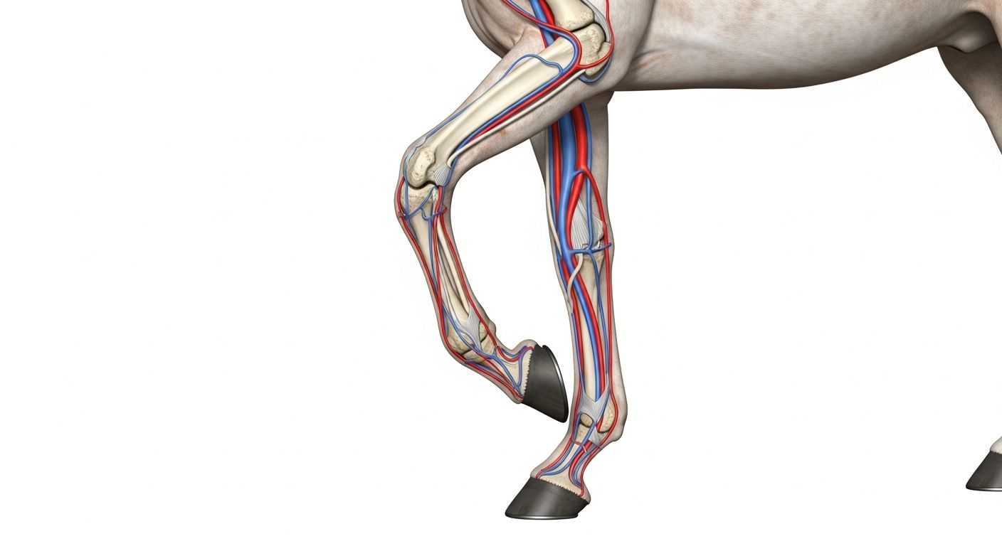
What Is the Distal Front Limb? A Suspension Bridge with No Maintenance Crew
Let’s walk it down—literally. The distal front limb (a core slice of the distal limb anatomy horse) is a miracle of minimalism:
| Structure | Function | “If It Fails…” |
|---|---|---|
| Carpus (8 small bones) | Shock absorption, directional control | Chip fractures → chronic synovitis |
| Metacarpus III (cannon) | Sole weight-bearing column below knee | Stress cracks if too fine or overworked |
| Proximal phalanx (P1 / long pastern) | Lever for breakover, joint stability | Flaring → strain on suspensory |
| Middle phalanx (P2 / short pastern) | Navicular support, angle control | Rotation → navicular syndrome |
| Distal phalanx (P3 / coffin bone) | Keystone of hoof, laminae anchor | Tip rotation = laminitis emergency |
No redundancy. No spares. Just elegant, high-stakes physics. Respect the distal limb anatomy horse, or the ground’ll school ya—for £120/hour.
What Are the Parts of a Horse Limb? Spoiler: It’s Not “Leg, Knee, Hoof”
Let’s name the gang—properly. In the full distal limb anatomy horse, we’ve got three zones:
- Proximal (power zone): Scapula/humerus (front), pelvis/femur (hind)—*muscle-heavy*, for drive and swing.
- Mid (leverage zone): Elbow/stifle → carpus/tarsus—*joint-dense*, for shock and direction.
- Distal (precision zone): Cannon down—*tendon & bone only*, for speed and economy.
Key tendons? Superficial digital flexor (SDFT) stores 60% of elastic energy at gallop. Deep digital flexor (DDFT) controls P3 angle—tighten it, and you pinch the navicular. Check ligaments (superficial & deep) are the “safety valves”—fail, and tendons overstretch. The distal limb anatomy horse isn’t built for *strength*—it’s built for *efficiency*. Every gram saved distally = less inertia = faster stride.
The Hoof Capsule: Where Distal Limb Anatomy Horse Meets the Earth
Frog, Sole, Wall—More Than Just Keratin
Down at ground zero, the hoof isn’t *dead*—it’s *dynamic*. In the distal limb anatomy horse, the capsule works like a heart:
- Frog: compresses on impact → pumps blood from digital cushion up the leg.
- Heel bulbs: expand laterally → absorb concussion.
- White line: the “seal” between wall and sole—stretch it, and bacteria waltz in (hello, abscess).
- Laminae: 600+ interlocking folds—bond P3 to wall. Fail? P3 rotates. *Disaster*.
A horse’s hoof hits the ground ~25,000 times/day in turnout. That’s why the distal limb anatomy horse *must* load evenly—or one structure bears the brunt. And nature *hates* imbalance.
Blood Flow & the Hoof Mechanism: Nature’s Best-Kept Secret
Here’s the magic most miss: the hoof *pumps blood*. Every stride:
- Heel lands → frog compresses → digital cushion flattens.
- Venous plexuses around P3 squeeze → blood pushed proximally.
- Hoof lifts → elastic recoil → vacuum effect → fresh arterial blood floods in.
This “hoof mechanism” contributes ~30% of venous return from the limb. Stall-bound horses? Poor circulation → colder feet → higher laminitis risk. That’s why turnout isn’t *nice*—it’s *non-negotiable*. The distal limb anatomy horse thrives on *movement*, not rest.
Common Injuries—And Where They Show Up in the Diagram
Before the limp, before the heat—the distal limb anatomy horse whispers:
| Sign | Likely Structure Involved | Urgency |
|---|---|---|
| Heat/swelling mid-cannon | SDFT injury (bowed tendon) | 🔴 Immediate vet |
| Lameness worse on hard circles | Navicular inflammation / DDFT strain | 🟡 48h window |
| Toe-first landing + short stride | Heel pain (thrush, underrun, bruising) | 🟢 Assess farrier |
| Asymmetrical sweat, cold back | Compensatory strain from distal imbalance | 🟡 Full biomechanical eval |
Early detection = cheap rehab. Late? £2,000+ and 6 months box rest. The distal limb anatomy horse doesn’t bluff—it *adapts*, silently, until it can’t.
Farriery & Trimming: Reading the Distal Limb Like a Weather Forecast
A trim isn’t *cosmetic*—it’s *biomechanical recalibration*. In the distal limb anatomy horse, key goals:
- Breakover point: should sit just in front of P3 tip. Too far forward? DDFT strain. Too far back? Tripping.
- Heel support: must sit *under* the widest part of the frog—not pinched, not floating.
- Medial-lateral balance: hoof-pastern axis aligned—not “perfect”, but *functional for that conformation*.
Shoes? Just *tools*. Heart bar → frog support. Rolled toe → ease breakover. But the best shoe’s the one that *lets the hoof work as evolution designed it*—in the distal limb anatomy horse, less is often more.
Conditioning the Distal Limb: Not Just “More Work”—*Smarter* Work
Tendons adapt *slower* than muscle—collagen turnover takes ~200 days. So “just lunge him more” is a fast track to injury. Smart conditioning for the distal limb anatomy horse:
- Walking on varied terrain (gravel, sand, grass) → stimulates sole thickening & frog callus.
- Trot poles (low, wide spacing) → encourages heel-first landing, activates digital cushion.
- Hillwork (shallow incline) → builds deep stabilisers without concussion.
- 24h turnout minimum → maintains hoof mechanism, circulation, proprioception.
Remember: the distal limb has *no muscles*—so strength comes from *tendon resilience*, *ligament integrity*, and *perfect alignment*. Train the *whole horse*, or the distal limb anatomy horse pays the price.
Where to Go Next: Your Distal Limb Mastery Starts Here
Ready to go deeper than the keratin? Start at Riding London for the big picture, nip into our Learn hub for guided walkthroughs (no vet degree required), or—if full-body context helps—grab a brew and explore parts to a horse labeled body parts guide. There’s 3D rotatable models, palpation guides, even a “what this means for your stride” decoder. Because the distal limb anatomy horse isn’t just for farriers and vets—it’s for *anyone* who’s ever whispered, *“How do you carry me on *that*?”* and meant it with awe.
FAQ
What is the distal limb of the horse?
The distal limb of the horse refers to the portion from the carpus (forelimb “knee”) or tarsus (hindlimb “hock”) down to the ground—encompassing cannon bone, pasterns, coffin bone, tendons, ligaments, and hoof capsule. In the distal limb anatomy horse, this region contains *no muscles*; movement is driven by tendons originating proximally. It functions as a high-efficiency, shock-absorbing lever system optimised for speed and endurance.
What is proximal and distal on a horse?
In equine anatomy, *proximal* means closer to the body’s core (e.g., shoulder, stifle), while *distal* means farther from the core (e.g., hoof, pastern). For example, the fetlock is *distal* to the knee, and the coffin bone is the *most distal* bone in the limb. Understanding this directional language is essential when interpreting the distal limb anatomy horse and communicating with vets or farriers.
What is the distal front limb?
The distal front limb is the section from the carpus (“knee”) down, including: *metacarpus III* (cannon bone), *proximal phalanx* (long pastern), *middle phalanx* (short pastern), *distal phalanx* (coffin bone), navicular bone, digital cushion, and hoof structures. In the distal limb anatomy horse, it’s a tendon-driven, muscle-free lever designed for elastic energy storage, shock absorption, and precise ground interaction.
What are the parts of a horse limb?
A horse’s limb is divided into: *proximal* (scapula/humerus or pelvis/femur—muscle-rich for power), *mid* (elbow/stifle to carpus/tarsus—joint-dense for shock control), and *distal* (cannon to hoof—tendon-and-bone only for efficiency). Key structures include bones (e.g. MCIII, P1–P3), tendons (SDFT, DDFT), ligaments (check ligaments, suspensory), and the hoof capsule. The distal limb anatomy horse highlights how evolution stripped away distal muscle to maximise speed and reduce fatigue.
References
- https://www.ncbi.nlm.nih.gov/pmc/articles/PMC7294591/
- https://www.rvc.ac.uk/research/research-centres/locomotion/equine-distal-limb-biomechanics
- https://www.vetfolio.com/manage/equine-lameness-differential-diagnosis-distal-limb
- https://www.sciencedirect.com/science/article/pii/S0737080622000746