Anatomy Hoof Horse Simplified Structure
- 1.
What is the anatomy of the horse hoof? Let’s pop the bonnet—this ain’t just a keratin cap, innit
- 2.
What is the middle part of a horse's hoof called? Meet the frog—no croaking, but plenty of squelch
- 3.
How should a horse's hoof look? Picture-perfect isn’t vanity—it’s physics with manners
- 4.
What's the sensitive tissue underneath the hoof that attaches to the bone of the toe? Laminae—nature’s Velcro, wired to a siren
- 5.
The hoof wall: laminated keratin, grown fresh like Sunday papers
- 6.
The sole: not for walking on—more like the lid on a treasure chest
- 7.
The digital cushion: padding with purpose, like memory foam in your nan’s slippers
- 8.
Hoof balance stats—because numbers don’t lie (even if your horse does)
- 9.
Hoof care myths—busted like a dodgy firework on Bonfire Night
- 10.
Where to go next? Keep the kettle on—we’re just getting started
Table of Contents
anatomy hoof horse
What is the anatomy of the horse hoof? Let’s pop the bonnet—this ain’t just a keratin cap, innit
Right—ever looked at a horse’s foot and thought, “Blimey, that’s just a big toenail glued to a boot?” Oh, *dear heart*. The anatomy hoof horse is more layered than a proper Bakewell tart—crust, jam, frangipane, *and* flaky top. Outer wall? Tough as a docker’s handshake. Sole? Concave shield—not for stepping on, *for* protecting. Frog? That squishy V? It’s a shock absorber, circulatory pump, *and* traction pad—all in one. Inside? Coffin bone (P3), navicular, digital cushion, and the *laminae*—those feathery tissues that bind bone to wall like a zip on a Barbour. Mess with one bit, and the whole symphony wobbles. A proper anatomy hoof horse diagram doesn’t just label—it *conducts* function, force, and fragility in one sweep.
What is the middle part of a horse's hoof called? Meet the frog—no croaking, but plenty of squelch
Centre stage, rubbery, slightly groovy—literally—is the frog. Not “pad”, not “sponge”, not “hoof jelly” (though we *have* heard that down the yard). It’s the middle part of a horse's hoof, shaped like a heart that’s been gently folded. Vital? Absolutely. When the hoof hits the ground, the frog compresses, pushing blood *up* the leg—that’s the *hoof pump mechanism*, luv. Thin, shrunken, or thrushy? Circulation suffers, heels collapse, and your nag starts walking like he’s stepped on a Lego brick. In any decent anatomy hoof horse cross-section, the frog’s shaded darker—often pink or red—to flag its biomechanical *oomph*.
How should a horse's hoof look? Picture-perfect isn’t vanity—it’s physics with manners
A healthy hoof’s got symmetry sharper than a Savile Row chalk line. Front-on? Straight down the midline—no flares, no shears. Side-on? The hoof-pastern axis should flow *smooth*—like trains on parallel tracks. Sole? Concave—not flat like a stale crisp. Frog? Plump, elastic, split cleanly down the middle. Heels? Neither underrun (caved in) nor overgrown (sticking out like sore thumbs). Wall? Smooth, unbroken, growth rings even as a well-laid brick path. A proper anatomy hoof horse reference overlays angles: ideal toe angle 50–55°, matching pastern. Deviate? You’re borrowing trouble—ringbone, navicular, tendon strain. Think of it like tea: the right balance *matters*—too strong, too weak, and no one’s happy.
What's the sensitive tissue underneath the hoof that attaches to the bone of the toe? Laminae—nature’s Velcro, wired to a siren
Here’s where things get *spicy*. Tucked between coffin bone (P3) and inner wall are the laminae—two sets: *insensitive* (wall side) and *sensitive* (bone side). They interlock like teeth on a zip, but *each sensitive lamina* is studded with nerves and capillaries. So what's the sensitive tissue underneath the hoof that attaches to the bone of the toe? It’s the *sensitive laminae*—and they’re *exquisitely* reactive. Inflame them (laminitis), and your horse’s in agony—not from surface pain, but from *structural betrayal*. That’s why a bounding digital pulse or reluctance to turn *matters*—it’s the laminae screaming. Any authoritative anatomy hoof horse schematic highlights this zone in fine, feathery detail—because without lamellar integrity? No soundness. No ride. Just suffering.
The hoof wall: laminated keratin, grown fresh like Sunday papers
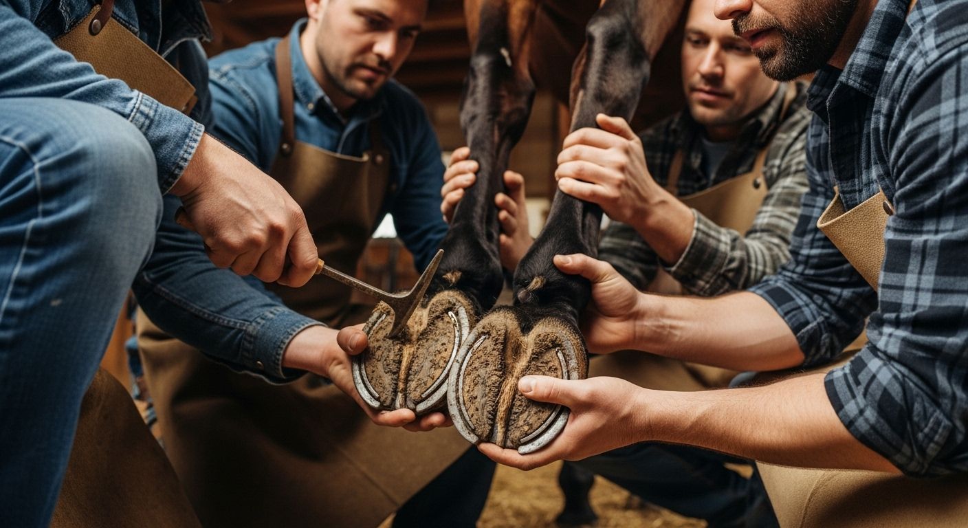
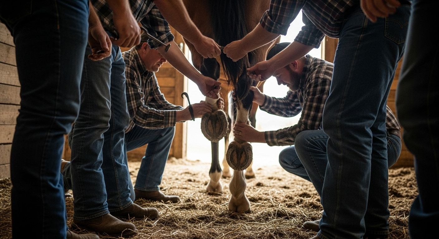
Ah, the hoof wall—nature’s carbon-fibre composite, laid down in tubules like terraced cottages on a Welsh hillside. It grows ~1 cm/month (faster in summer, slower in winter—just like your will to mow the lawn). It’s split into *toe*, *quarters*, and *heels*—each with its own job. Toe takes impact, quarters flex, heels expand. Cracks? Flares? Seedy toe? All red flags—often from imbalance, poor diet (low zinc, biotin), or infrequent farrier visits (£45–£75 every 6–8 weeks, mind you—worth every quid). In a crisp anatomy hoof horse illustration, the wall’s the bold outer rim—thick, resilient, *alive at the top*. But remember: growth without balance is just *more mess*, not more strength.
The sole: not for walking on—more like the lid on a treasure chest
Let’s clear the fog: the sole isn’t weight-bearing—it’s *protective*. Think of it as the hull of a boat: slightly concave, calloused, shielding the coffin bone and sole corium underneath. Pare it too thin (looking at you, overzealous hoof-pickers), and you expose sensitive tissue. Result? Lameness, bruising, “sore-footed” gait. Healthy sole is firm but flexible—not chalky, not flaky. In muddy paddocks? It softens. On dry, stony ground? It hardens. That’s *adaptation*, not weakness. A top-tier anatomy hoof horse diagram shades the sole *distinctly* from frog and wall—because confusing them is how beginners turn routine trims into vet emergencies.
The digital cushion: padding with purpose, like memory foam in your nan’s slippers
Tucked above the frog—soft, fibro-fatty, criminally underrated—is the digital cushion. It’s the hoof’s airbag. At impact, it spreads, flattens, and pushes the heels *outward*, helping the whole capsule expand. Foals? Mostly fat. Adults? Fibrous tissue takes over—*especially* in barefoot horses with proper ground contact. Shod too long? Cushion atrophies. Heels cave. Navicular strain creeps in. A good anatomy hoof horse cross-section shows it as a pale, wedge-shaped zone—often labelled “DC”—because without it, the hoof’s like a crisp packet with no crunch: all form, no function. Respect it, and your horse’ll move like poetry. Ignore it? Well… let’s just say the vet’s invoice’ll rhyme with “grim”.
Hoof balance stats—because numbers don’t lie (even if your horse does)
Let’s get clinical—for a sec. BEVA 2024 UK survey on hoof imbalances & lameness:
| Imbalance | Prevalence | Lameness Risk (vs. balanced) | Avg. Rehab Cost (GBP) |
|---|---|---|---|
| Underrun heels | 42% | 3.2× higher | £1,200 |
| Long toe / low heel | 38% | 2.8× higher | £950 |
| Medial-lateral asymmetry | 29% | 4.1× higher | £1,800 |
| Thin sole (<10mm) | 33% | 2.5× higher | £720 |
“An ounce of prevention’s worth a stone of cure,” as Nan used to say—especially when it comes to hoof health. Keep that anatomy hoof horse chart on your phone. Use it like a pub quiz cheat sheet: if something looks off, *act*. Time + ignorance = expensive, heart-breaking rehab.
Hoof care myths—busted like a dodgy firework on Bonfire Night
Let’s pop a few bubbles, shall we?
- “Hooves need shoes to stay strong.” → Nay. Many thrive barefoot—with movement, proper trim, and diet.
- “The white line’s supposed to be white.” → Nope. It’s yellowish-beige. “White” is old anatomist slang.
- “Frog doesn’t need ground contact.” → Wrong. No contact = atrophy = poor pump = cold legs.
- “Cracks are just cosmetic.” → Only if superficial. Deep cracks? Lameness waiting to happen.
Myths stick like mud on wellies—but a solid grasp of anatomy hoof horse clears the fog. Because knowledge? That’s the first step toward partnership—not just management.
Where to go next? Keep the kettle on—we’re just getting started
Fancy more? We’ve been down this rabbit hole for years—and we *love* it. Start at the source: Riding London, our humble homebase. Fancy structured learning? The Learn section’s got guides, quizzes, and proper nerd-outs for every level—from first-timers to seasoned yard hands. And if you *really* want to geek out on heritage and horsemanship beyond anatomy, don’t miss our deep-dive into legacy and lineage—reign horses: royal lineage equestrian brand. Because horses aren’t just biology—they’re history, culture, and heart, all in one gallop.
Frequently Asked Questions
What is the anatomy of the horse hoof?
The anatomy hoof horse includes external structures—the *hoof wall* (toe, quarters, heels), *sole*, *frog*, *bars*, and *white line*—and internal components: the *coffin bone (P3)*, *navicular bone*, *digital cushion*, *laminae* (sensitive + insensitive), and vascular networks. It functions as a dynamic shock absorber, circulatory pump, and weight-bearing platform. A detailed anatomy hoof horse diagram integrates all layers to show structural and functional interdependence.
What is the middle part of a horse's hoof called?
The middle part of a horse's hoof is called the *frog*—a wedge-shaped, elastic, V-shaped structure vital for shock absorption, traction, and activating the hoof pump mechanism. It is rich in nerve endings and sweat glands. In standard anatomy hoof horse illustrations, the frog appears centrally, often shaded distinctly to highlight its functional role in biomechanics and circulation.
How should a horse's hoof look?
A healthy hoof should be symmetrical, with a smooth, unbroken wall, no flaring or cracks. The sole is concave—not flat—and the frog is wide, elastic, and centrally grooved. Heels should be neither underrun nor overgrown, and the hoof-pastern axis (side view) aligns straight. Any deviation warns of imbalance. Cross-referencing with a reliable anatomy hoof horse guide helps spot subtler issues before they worsen into lameness or pathology.
What's the sensitive tissue underneath the hoof that attaches to the bone of the toe?
What's the sensitive tissue underneath the hoof that attaches to the bone of the toe? It’s the *sensitive laminae*—the vascular, highly innervated tissue that interlocks with the insensitive laminae of the hoof wall to secure the coffin bone (P3). This junction is critical for structural stability and is the primary site affected in laminitis. Understanding this in the context of anatomy hoof horse is essential for early detection and prevention of catastrophic hoof failure.
References
- https://www.ncbi.nlm.nih.gov/pmc/articles/PMC6383295
- https://www.bevalibrary.org/horse-hoof-anatomy
- https://courses.ecampus.oregonstate.edu/equine/human-horse-relationship/hoof-anatomy